
30 Winning Shots From Nikon’s Small World Photomicrography Competition 2022
This is the 48th year that Nikon invited photographers to its Small World Photomicrography Competition, and there are so many amazing entries from all around the globe this year.
The awards have been announced and the winning images take us to the wonderful and surprising microscopic world. Many scientists and photography enthusiasts peeped into the light microscope to show us the amazing things that we cannot see with our naked eyes.
Check out some of the most stunning images that won this year in the gallery below.
More info: nikonsmallworld.com | Instagram | Facebook | twitter.com
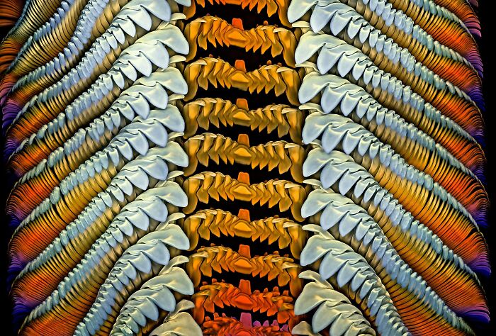
#1 1st Place – Grigorii Timin, Dr. Michel Milinkovitch

“Embryonic hand of a Madagascar giant day gecko (Phelsuma grandis).”
University of Geneva, Geneva, Switzerland Department of Genetics and Evolution
#2 2nd Place-Caleb Dawson

“Breast tissue showing contractile myoepithelial cells wrapped around milk-producing alveoli”
WEHI, The Walter and Eliza Hall Institute of Medical Research Department of Immunology Melbourne, Victoria, Australia
#3 3rd Place Satu PaavonsaloDr. Sinem Karaman

“Blood vessel networks in the intestine of an adult mouse”
University of Helsinki Individualized Drug Therapy Research Program, Faculty of Medicine Helsinki, Finland
#4 4th Place – Dr. Andrew Posselt

“Long-bodied cellar/daddy long-legs spider (Pholcus phalangioides).”
University of California, San Francisco (UCSF), Mill Valley, California, USA Department of Surgery
#5 5th Place – Alison Pollack

“Slime mold (Lamproderma).”
San Anselmo, California, USA
#6 Honorable Mention – Ye Fei Zhang

“Butterfly egg.”
Jiang Yin, Jiangsu, China
#7 Image Of Distinction – Xinpei Zhang

“Alaskan sand.”
Yu Cheng, Ya’an, China
#8 Image Of Distinction – Dr. Andrew Posselt

“Bold jumping spider (Phidippus audax).”
University of California, San Francisco (UCSF) Department of Surgery Mill Valley, California, USA
#9 Image Of Distinction – Karl Deckart

“Dental drill bit studded with diamond chips.”
Eckental, Bavaria, Germany
#10 Image Of Distinction – Gabriel Fernández Fernández Jorge Alberto

“Four o’clock flower (Mirabilis jalapa).”
San Luis, Argentina
#11 Image Of Distinction – Ahmad Fauzan

“Black and white human hair.”
Macro Depok (MD) Department of Engineering Jakarta, Indonesia
#12 Image Of Distinction – Dr. Eugenijus Kavaliauskas

“Ant (Camponotus).”
Tauragė, Lithuania
#13 Image Of Distinction – Anne-Françoise Tasnier

“Wood cells.”
Royal Museum for Central Africa Department of Wood Biology Tervuren, Belgium
#14 Image Of Distinction – Yuan Ji

“Butterfly scales.”
World Expo Museum Shanghai, China
#15 Honorable Mention – Sebastian Sparenga

“Recrystallized Vitamin C.”
McCrone Research Institute Chicago, Illinois, USA
#16 10th Place – Murat Öztürk

“A fly under the chin of a tiger beetle.”
Ankara, Turkey
#17 Image Of Distinction – Adolfo Ruiz De Segovia

“Drops of olive oil in water.”
Particular Madrid, Spain
#18 6th Place – Ole Bielfeldt

“Unburned particles of carbon released when the hydrocarbon chain of candle wax breaks down.”
Macrofying Cologne, North Rhine-Westphalia, Germany
#19 Image Of Distinction – Yoshihiro Tamaru

“Tail of a planktonic crustacean (Oithona brevicornis).”
Hino, Tokyo, Japan
#20 Image Of Distinction – Michael Landgrebe

“Moss spore capsule (sporangium).”
Berlin, Germany
#21 Image Of Distinction – Teresa Zgoda

“Eyeshadow cosmetic.”
Arvada, Colorado, USA
#22 Image Of Distinction – Dr. Stephen S. Nagy

“Diatoms arranged in an exhibition rosette by Klaus D. Kemp.”
Montana Diatoms Helena, Montana, USA
#23 Image Of Distinction – Dr. Honor Glenn

“Human lung cell infected with coronavirus.”
Arizona State University Biodesign Institute Biodesign Imaging Facility, Center for Immunotherapy, Vaccines, and Virotherapy Tempe, Arizona, USA
#24 Image Of Distinction – Anatoly Mikhaltsov

“Cross section of a leaf of dune grass (Ammophila arenaria).”
Children’s Ecological and Biological Center Department of Botany Omsk, Russia
#25 Honorable Mention – Dr. Igor Siwanowicz
“Radula (rasping tongue) of a marine snail (Turbinidae family).”
Howard Hughes Medical Institute (HHMI), Ashburn, Virginia, USA Janelia Research Campus
#26 14th Place – Nadia Efimova

“Differentiated cultured mouse myoblasts with lysosomes (cyan/green), nuclei (yellow), F-actin (magenta).”
Amicus Therapeutics Philadelphia, Pennsylvania, USA
#27 8th Place – Dr. Nathanaël Prunet

“Growing tip of a red algae.”
University of North Carolina at Chapel Hill, Chapel Hill, North Carolina, USA Department of Biology
#28 Honorable Mention – Dr. Laurent Formery

“Two-month old juvenile sea star (Patiria miniata).”
University of California, Berkeley, Berkeley, California, USA Department of Molecular and Cell Biology
#29 11th Place – Ye Fei Zhang

“Moth eggs.”
Jiang Yin, Jiangsu, China
#30 Image Of Distinction – Dr. Marko Pende

“Transgenic axolotl (CNP:GFP;β3Tubulin:mCherry) showing components of the nervous system. CNP+ Schwann cells (cyan) and axons (magenta).”
MDI Biological Laboratory Bar Harbor, Maine, USA


Saumya Ratan
Saumya is an explorer of all things beautiful, quirky, and heartwarming. With her knack for art, design, photography, fun trivia, and internet humor, she takes you on a journey through the lighter side of pop culture.
