20 Spectacular Photomicrography Winners At The 2025 Nikon Small World Contest
Widely regarded as the leading contest for recognising the art, skill, and excellence in photomicrography, the Nikon Small World competition is now in its 51st year. This year, the 2025 Nikon Small World Photomicrography Competition received 1,925 photo entries from 77 countries, with 20 winners selected for their outstanding submissions. Each image highlights the beauty and complexity of subjects seen through a light microscope, revealing aspects of the world invisible to the naked eye. Through their unique perspectives, these photographers offer everyone a chance to witness the breathtaking wonders of the microscopic world.
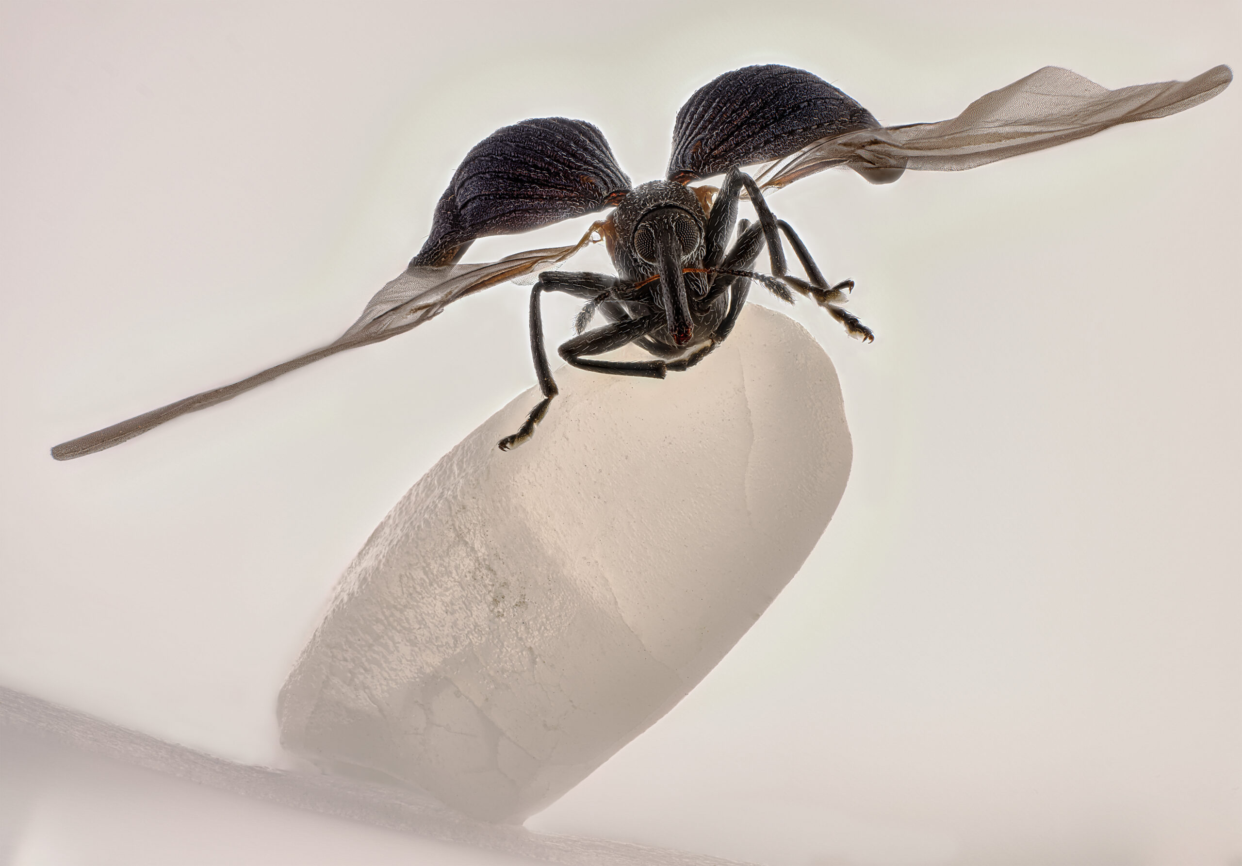
China’s Zhang You claimed the prestigious first place with his striking image of a rice weevil perched on a grain of rice. The photo captures the insect with its wings fully extended, frozen in a moment that showcases the form and behaviour of this common but often overlooked agricultural pest. “Zhang You’s work demonstrates the remarkable power of microscopy to reveal new perspectives on the world around us,” said Eric Flem, Senior Manager, Communications and CRM at Nikon Instruments. “What makes this year even more extraordinary is that it was his very first time entering the competition, and he not only captured first place, but also placed another image in the top 20. His achievement highlights the spirit of Nikon Small World: inspiring wonder, making scientific understanding accessible to all, and celebrating the artistry of the microscopic realm.
More info: Instagram | Facebook | LinkedIn | X
#1 1st place – You Zhang (Rice weevil (Sitophilus oryzae) on a grain of rice)
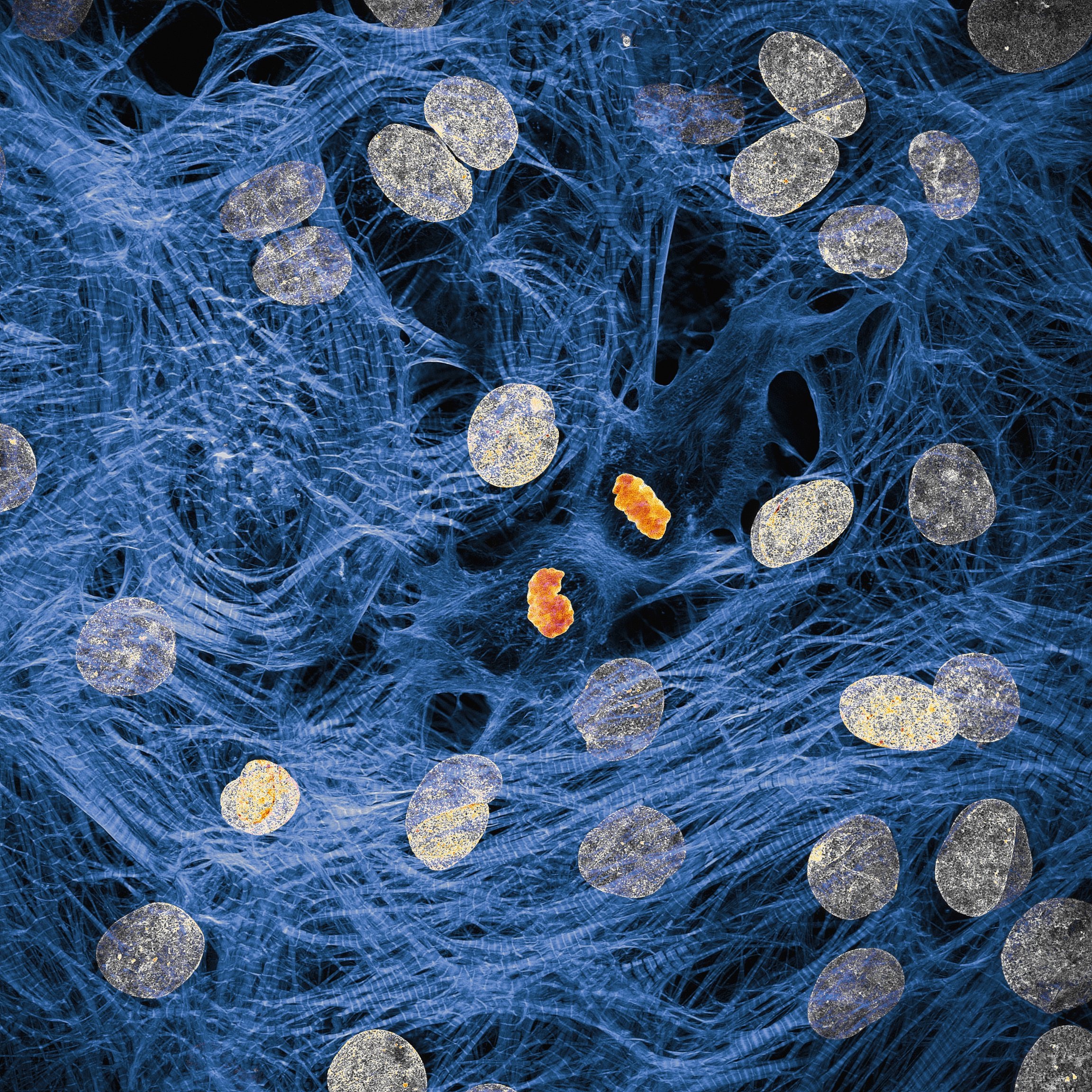
#2 6th place – Dr. Francisco Lázaro-Diéguez (Rat liver cells)
#3 13th place – Henri Koskinen (Slime mold (Arcyria major) releasing spores)
#4 4th place – Dr. James Hayes (Heart muscle cells with chromosomes condensed following cell division)
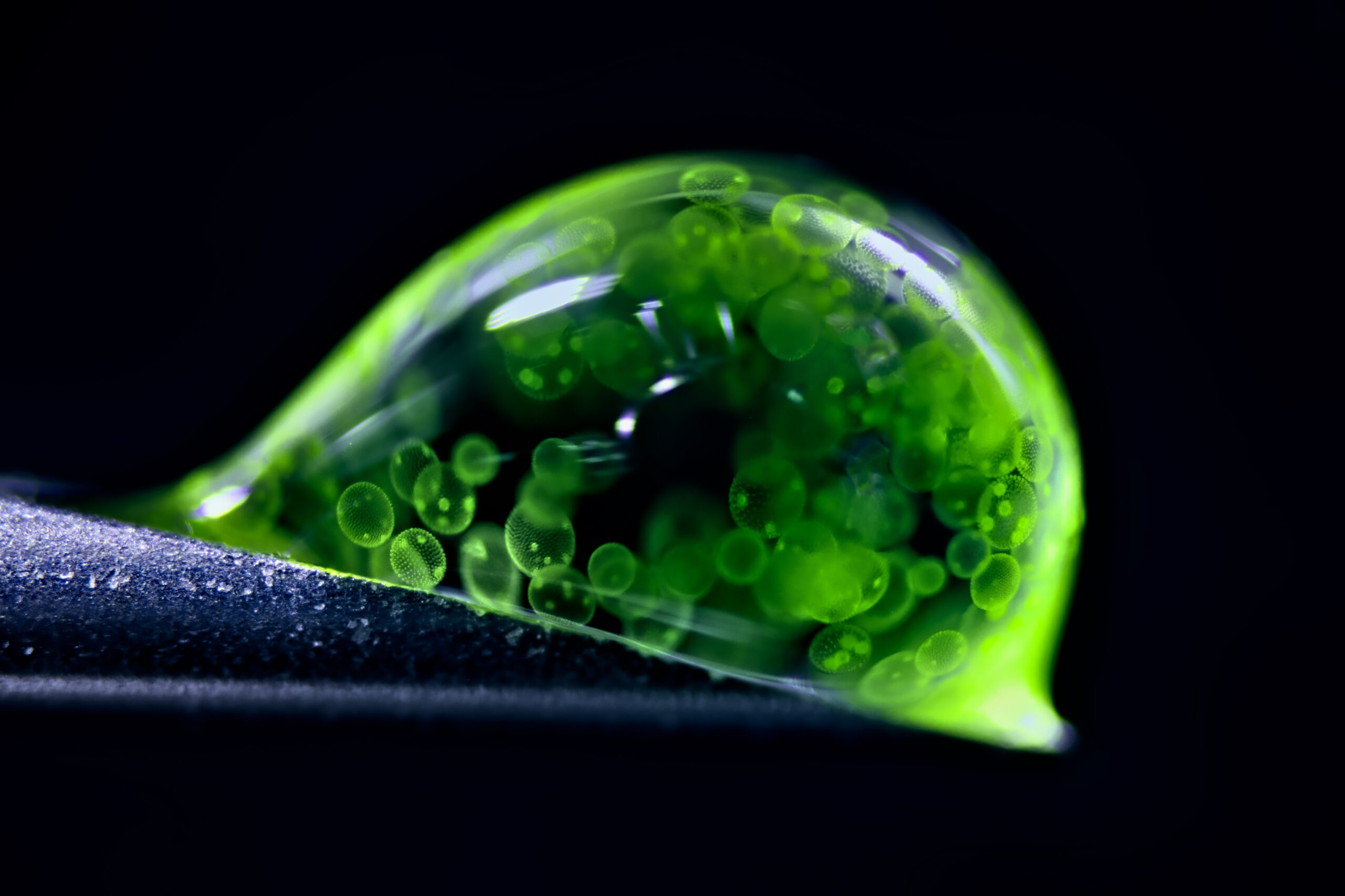
#5 2nd place – Dr. Jan Rosenboom (Colonial algae (Volvox) spheres in a drop of water)
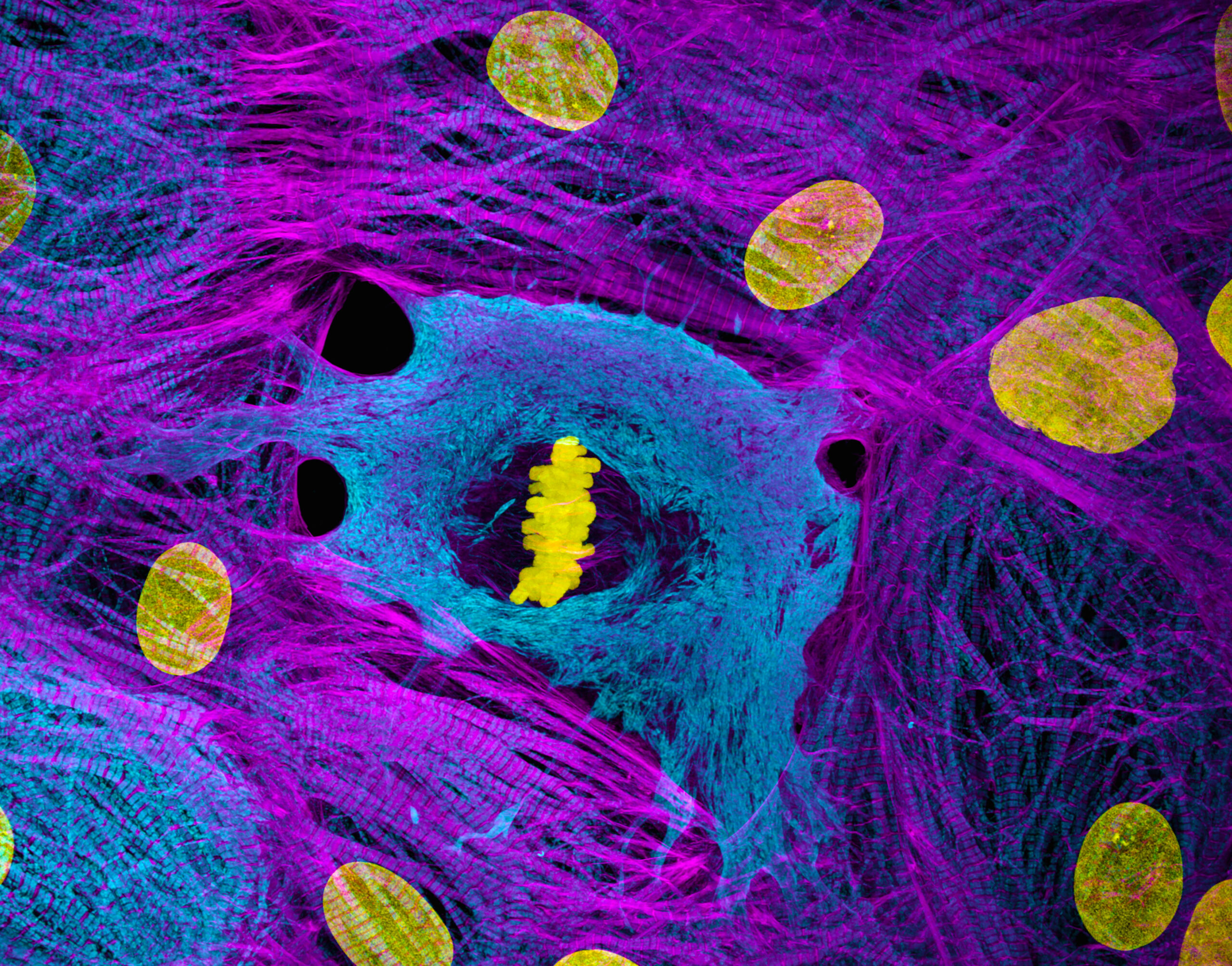
#6 10th place – Dr. Dylan Burnette (Heart muscle cells (iPSC-derived) showing condensed chromosomes in metaphase)
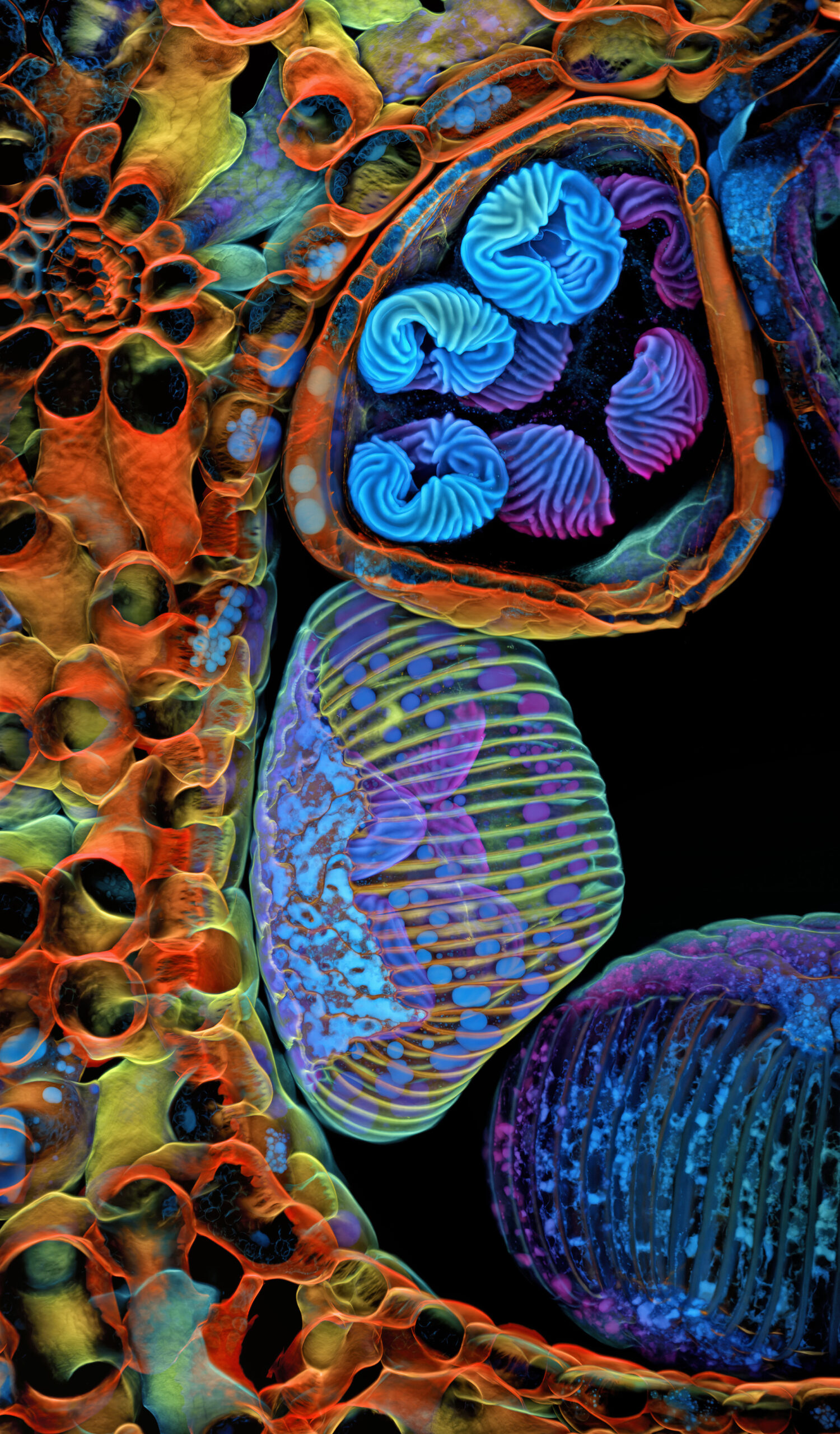
#7 5th place – Dr. Igor Siwanowicz Spores (blue/purple structures) of a small tropical fern (Ceratopteris richardii)
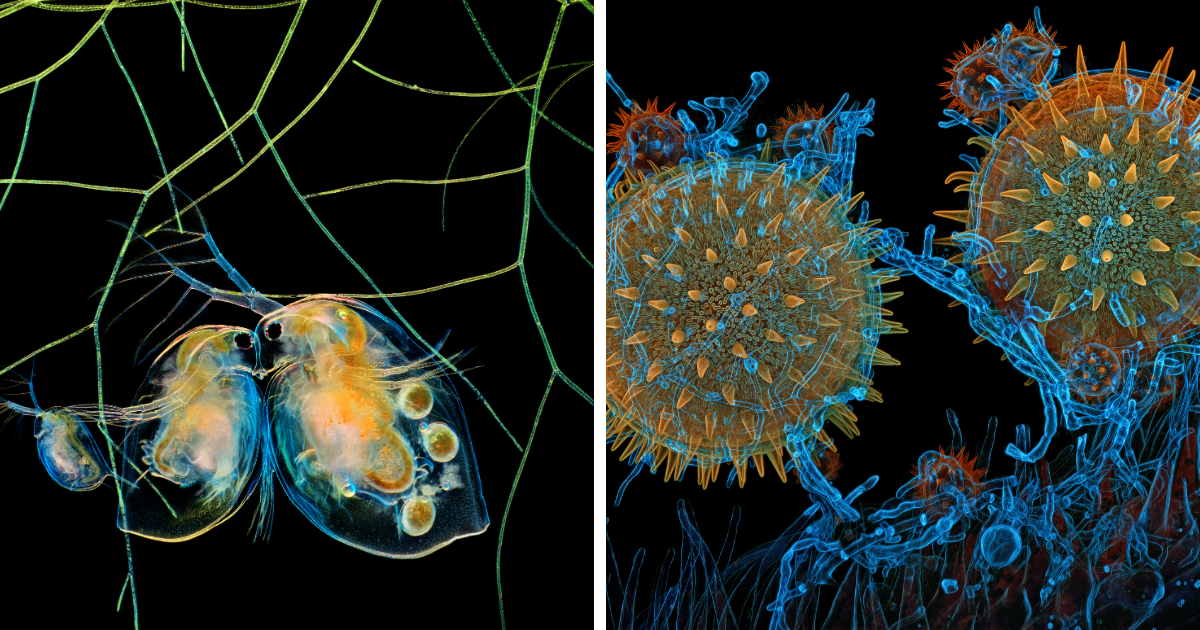
#8 3rd place – John-Oliver Dum (Pollen in a garden spider web)
#9 7th place – Stella Whittaker (iPSC-derived sensory neurons labelled to show tubulin and actin)
#10 8th place – Dr. Igor Siwanowicz (Mallow pollen germinating on stigma while being parasitized by a filamentous fungus)
#11 17th place – Hong Guo (Water fleas (Daphnia) and algae)
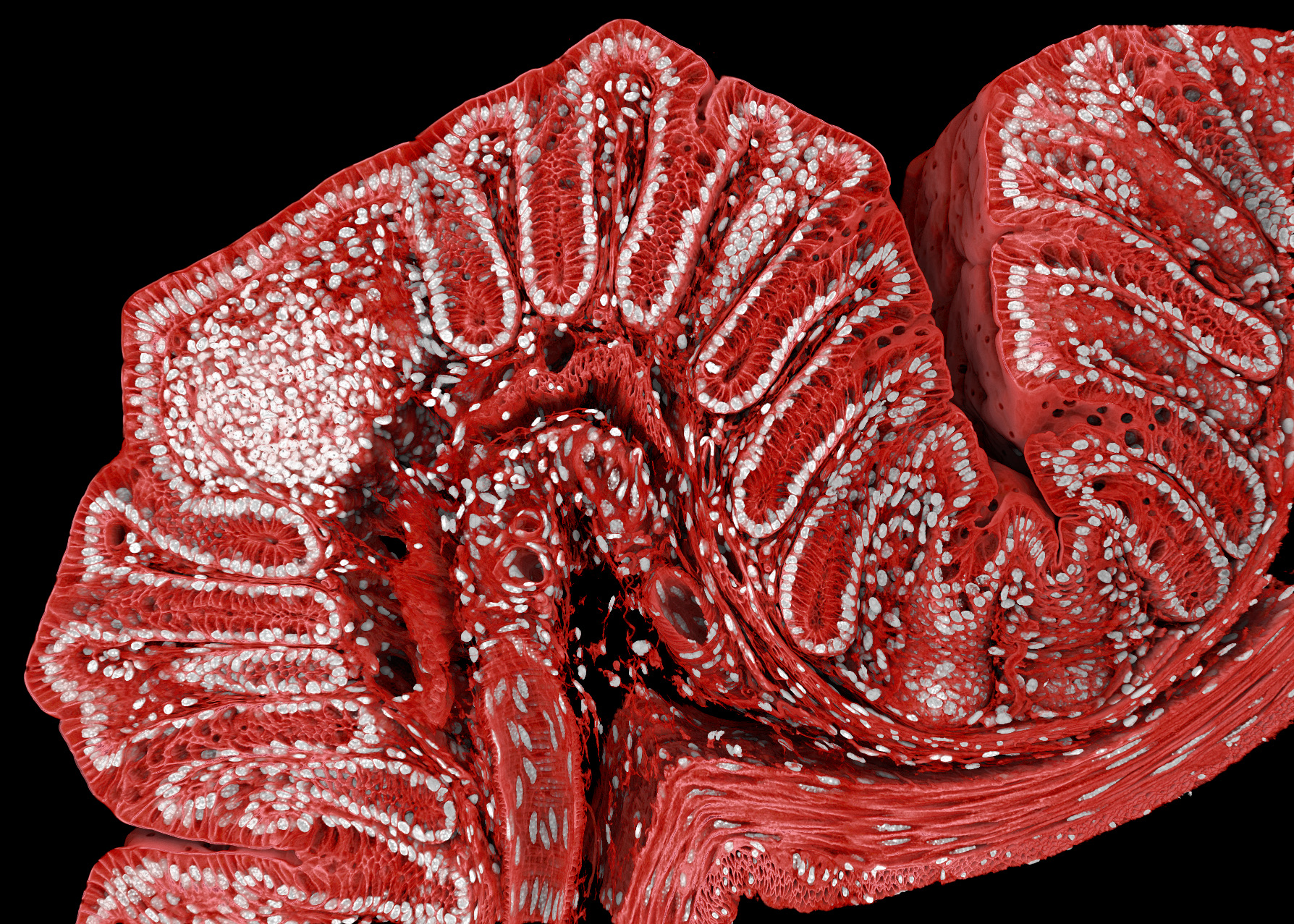
#12 18th place – Marius Mahlen (Fluorescently marked mouse colon)
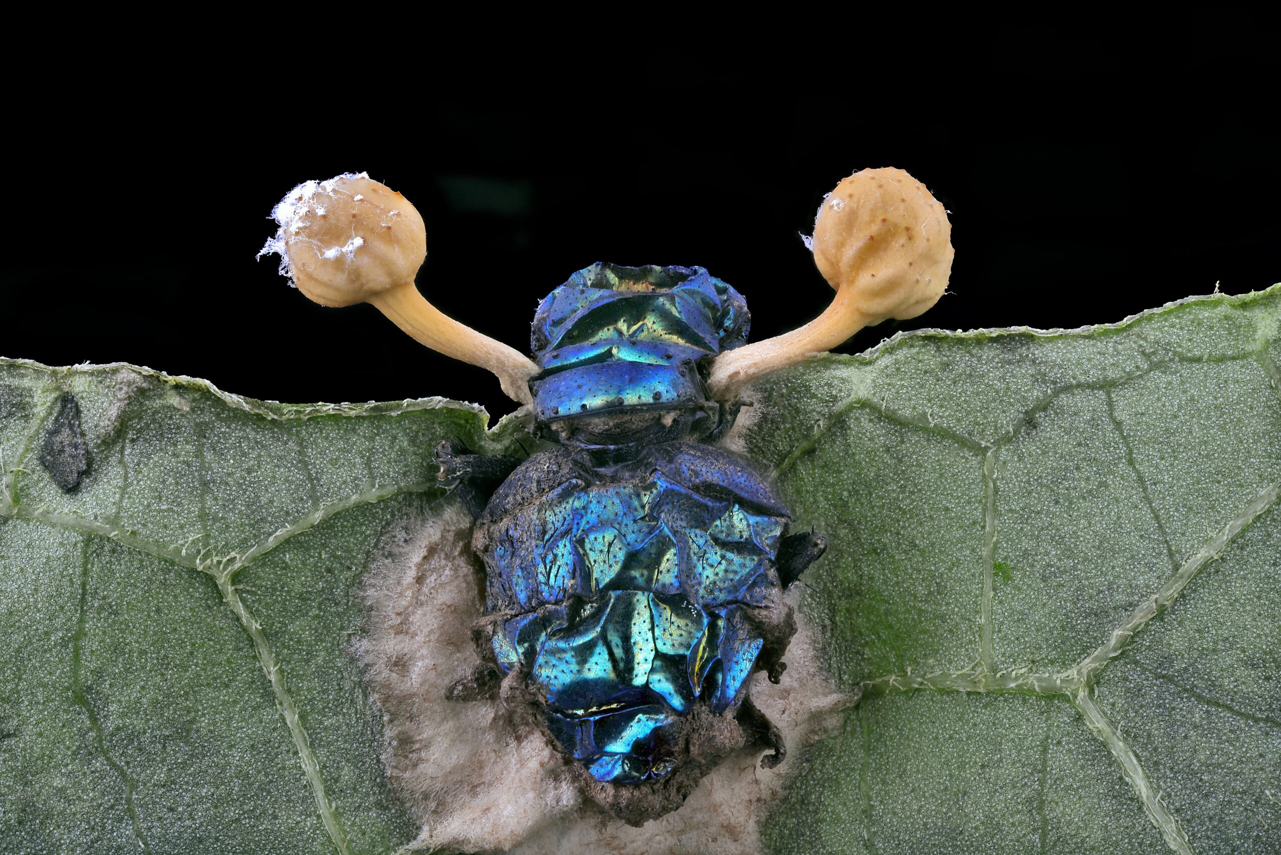
#13 19th place – Eduardo Carrasco (Parasitic fungus (Cordycipitaceae) on a fly (Calliphoridae))
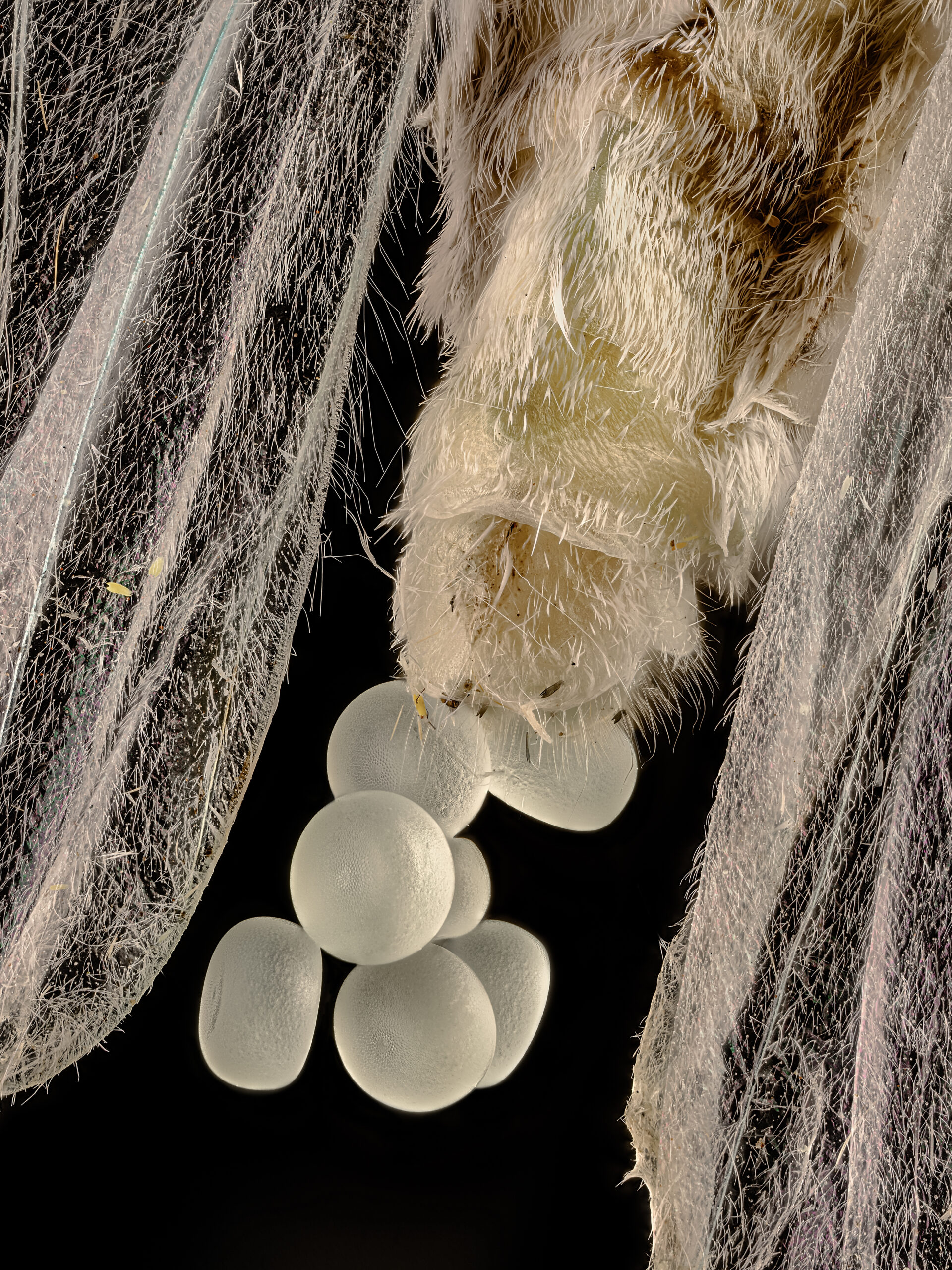
#14 15th place – You Zhang (Geometer moth (Geometridae) laying eggs)
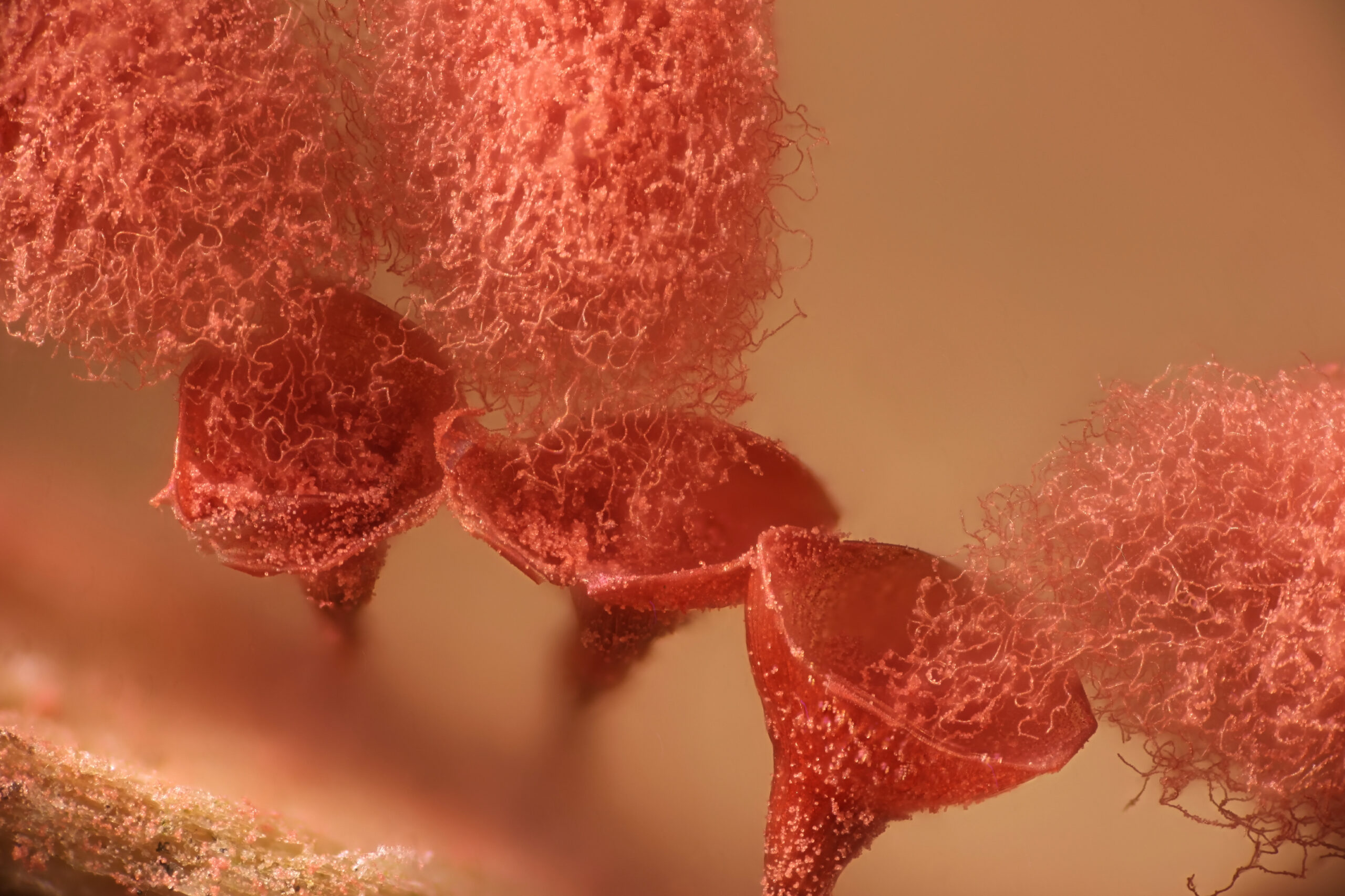
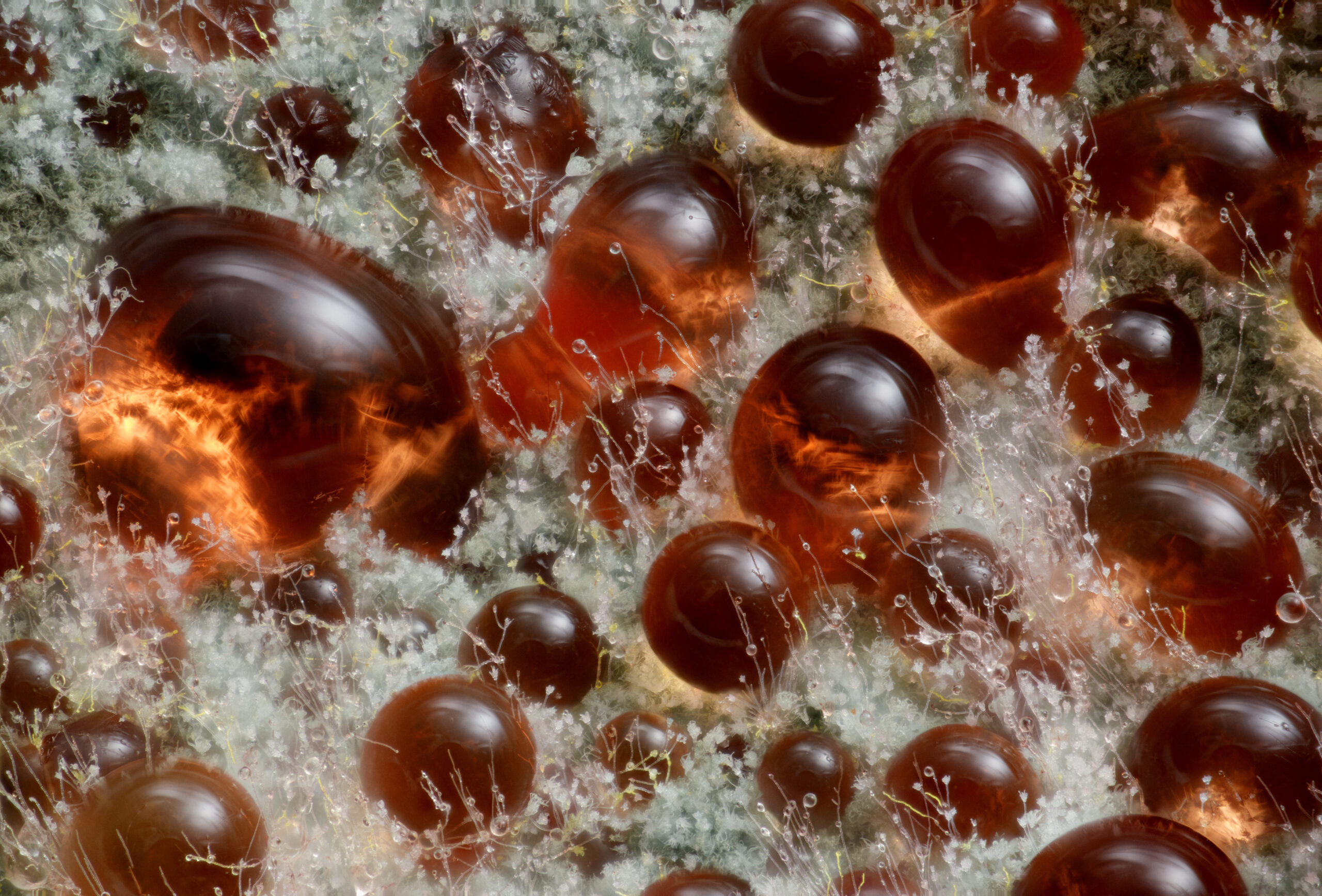
#15 9th place – Wim van Egmond (A fungus (Talaromyces purpureogenus) known for its red, diffused pigment)
#16 11th place – Marek Mis (Sunflower trichomes (hair-like plant outgrowths))
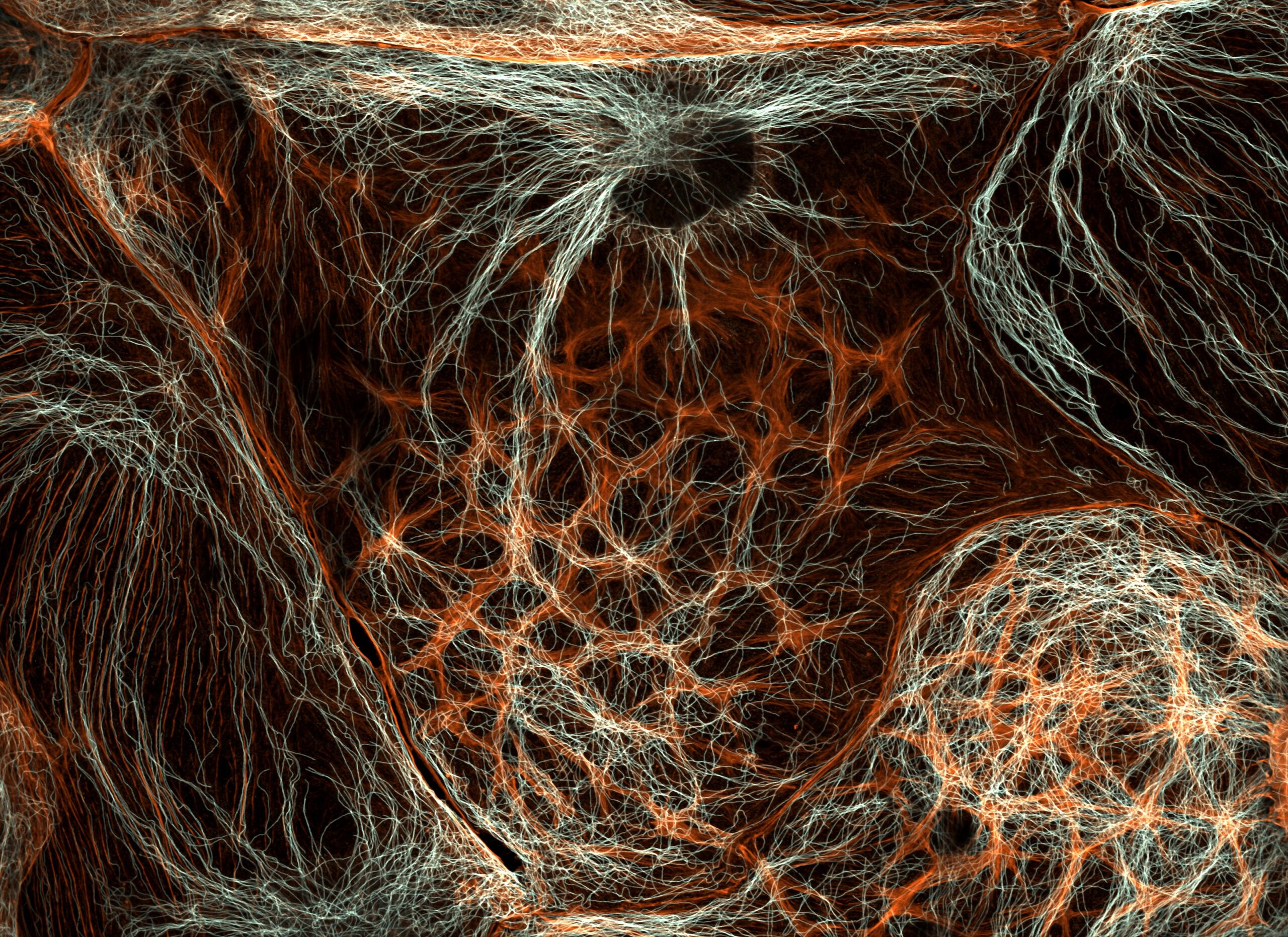
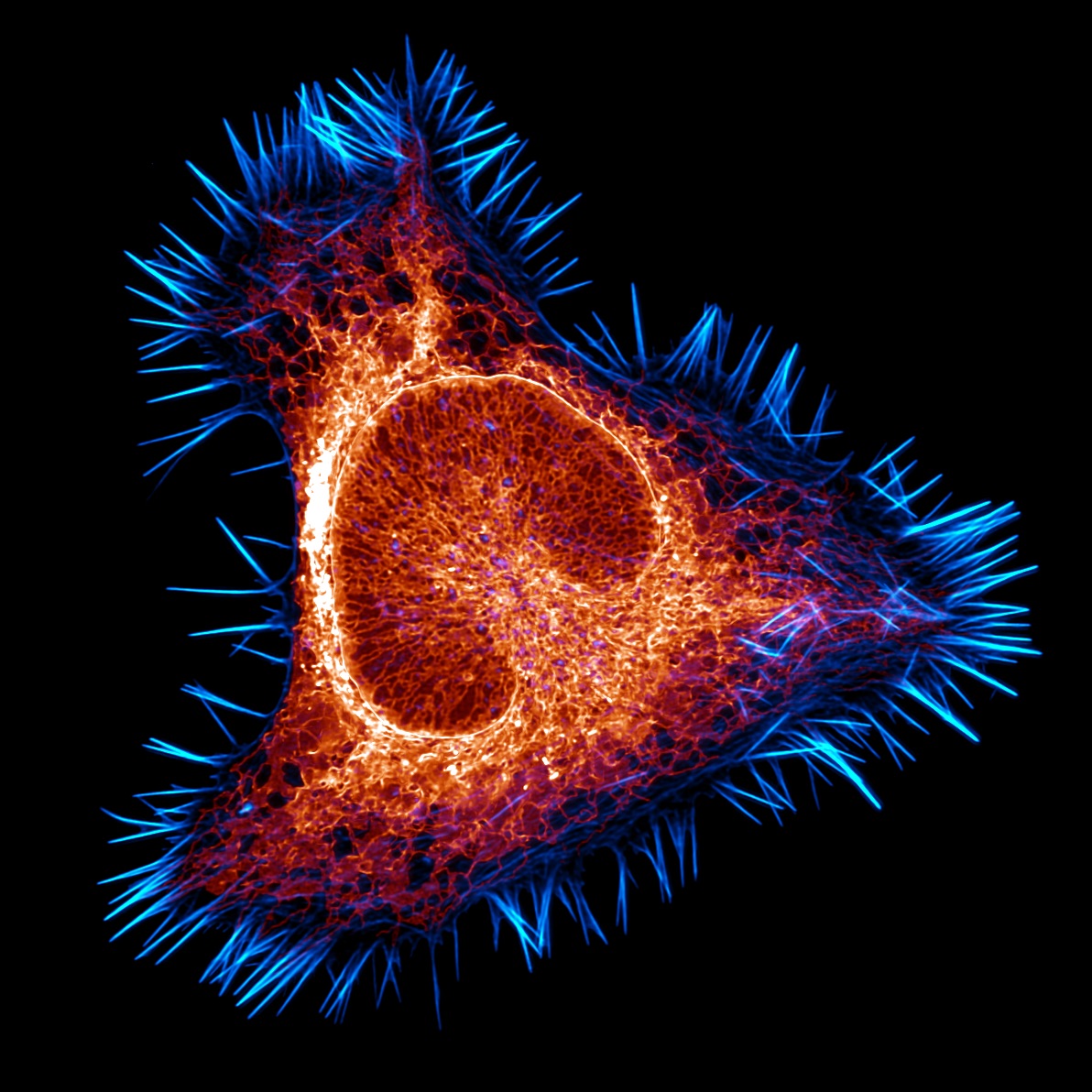
#17 12th place – Halli Lindamood (The actin cytoskeleton (cyan) and endoplasmic reticulum (red) of a mouse brain cancer cell)
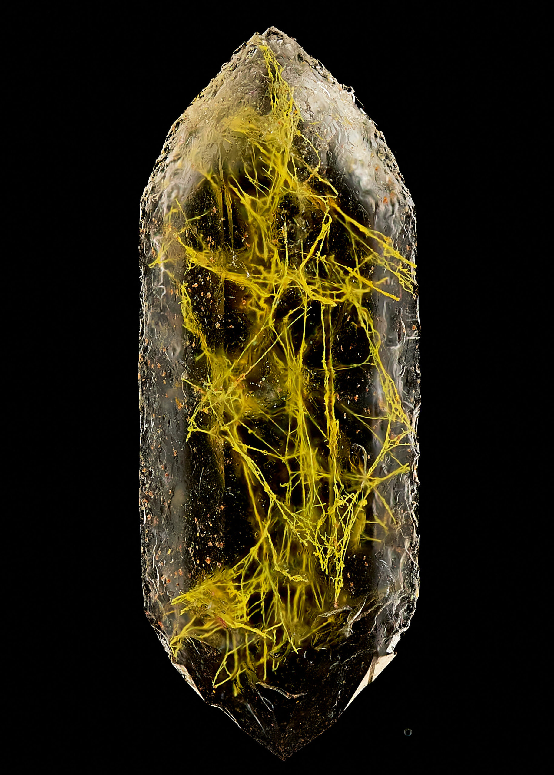
#18 14th place – Manfred Heising (Quartz with biotic goethite filaments)
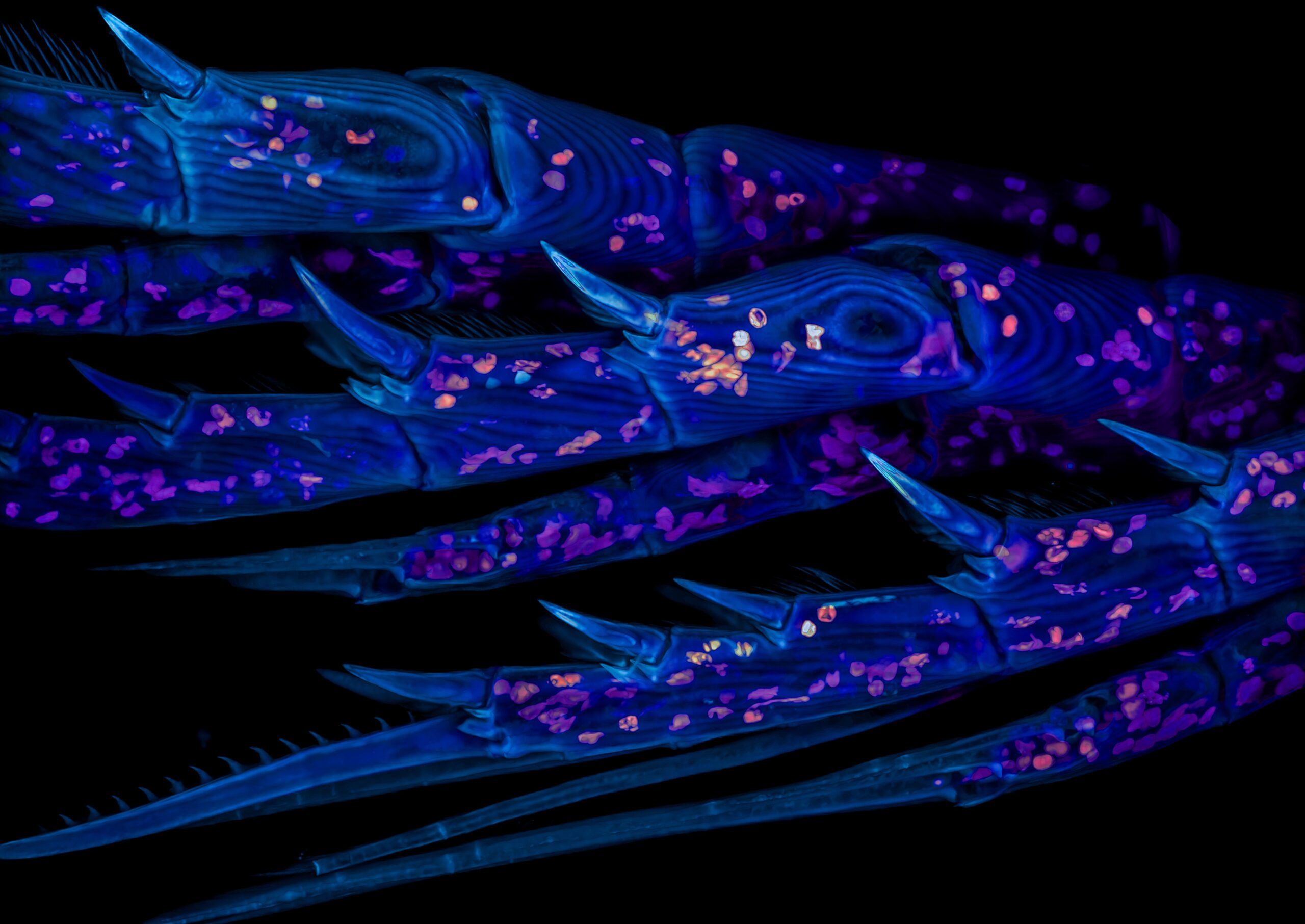
#19 20th place – Zachary Sanchez (Marine copepod)
#20 16th place – Rogelio Moreno (Spore sacs (sporangia) of a fern)


Shanilou Perera
Shanilou has always loved reading and learning about the world we live in. While she enjoys fictional books and stories just as much, since childhood she was especially fascinated by encyclopaedias and strangely enough, self-help books. As a kid, she spent most of her time consuming as much knowledge as she could get her hands on and could always be found at the library. Now, she still enjoys finding out about all the amazing things that surround us in our day-to-day lives and is blessed to be able to write about them to share with the whole world as a profession.
